High precision non-contact dispensing technologies and provides solutions for single cell analysis and cloning.
Accelerate to discover
Related topics

Spatial Proteomics: Charting the Protein Landscape in Tissues
What is Spatial Proteomics?
Spatial proteomics is the analysis of protein abundance, localization, and interactions within small tissue regions, specific cell populations, and subcellular compartments. While conventional proteomics analyzes large numbers of cells together, ignoring their heterogeneity and relying on averaged results, spatial proteomics offers a more accurate and detailed view of proteome dynamics and function. Crucially, it also enables the study of spatial relationships and molecular interactions between different regions or cell types within a tissue, shedding light on how local microenvironments and cell-to-cell communication shape biological processes.
The concept of microenvironments has gained significant attention, especially in key therapeutic fields such as oncology and immunology. Tumor cells interact with their environment differently from healthy cells, including with immune cells, which can either promote or inhibit tumor growth. Furthermore, tumors themselves commonly display distinct intra-tumor heterogeneity, where distinct cell populations engage in functional cross-talk within the same tumor. Spatial proteomics enables researchers to understand differences in cell behavior, ultimately providing insights into disease mechanisms that may lead to more targeted and effective treatment approaches.
Beyond assessments of heterogeneity, spatial proteomics also supports broader applications, such as immune response profiling, analysis of tissue morphology and microarchitecture, and the generation of high-resolution “atlases” of tissues and organs. These new capabilities have emerged thanks to recent technological advancements, including single-cell isolation and precise spatial sampling.
Technologies Driving Spatial Proteomics
Various technologies enhance resolution beyond that of conventional proteomic methods. Key spatial proteomics approaches include:
Imaging mass spectrometry
Imaging mass spectrometry (IMS) enables spatially resolved molecular analysis directly from tissue sections, providing “untargeted” maps of protein localization, that is, the analysis of a wide array of molecular species within a tissue sample without prior selection or labeling of specific targets. Matrix-Assisted Laser Desorption/Ionization IMS (MALDI-IMS) is the most commonly used technique in this category, allowing label-free detection of peptides and proteins across tissue sections. This methodology is particularly valuable in exploratory research, where the goal is to discover novel biomarkers or understand complex tissue heterogeneity without preconceived notions of which molecules are of interest.
Multiplexed antibody-based imaging
This technique utilizes sequential staining or specific antibodies labeled with fluorophores, metals, or DNA barcodes to simultaneously image dozens of protein targets in tissues, preserving spatial context. This ‘targeted’ method requires prior selection and validation of target proteins, making it more suitable for studies focusing on known markers or pathways.
Laser capture microdissection
This technique allows precise isolation of microscopic tissue regions or single cells under a microscope using laser targeting, enabling downstream proteomic analyses of targeted areas/populations. Laser capture microdissection (LMD) is typically performed on fixed tissues, commonly formalin-fixed paraffin-embedded (FFPE) samples, or frozen samples, providing structural stability that is crucial for precise microdissection.
Spatial barcoding workflows
Spatial barcoding workflows utilize techniques such as microfluidics or in situ barcoded arrays to analyze transcriptomes and proteins within intact tissue sections. For instance, DBiT-seq involves the sequential application of two sets of DNA barcodes through orthogonal microfluidic channels, creating a grid of uniquely barcoded pixels across the tissue. Antibodies are designed to specifically bind to target proteins, each antibody being conjugated to one of these unique DNA barcodes.
Deep visual proteomics
This technique combines high-resolution imaging, AI-guided cell selection, and ultra-sensitive mass spectrometry to quantify proteins in specific cells within complex tissues. For instance, it has been utilized to analyze archived tissue samples, such as primary melanoma tissues, to identify spatially resolved proteome changes during disease progression.
The Role of Single-Cell Sample Preparation
Researchers using spatial proteomics face challenges during sample preparation, particularly when using traditional techniques that lack the precision needed for accurately processing very small sample quantities. These challenges include:
Sample scarcity and low input
Spatial proteomics often targets minute regions of tissue or single cells, where available material is extremely limited. Conventional proteomic workflows typically require bulk input and may not be optimized for low-abundance samples, leading to incomplete or biased protein detection.
Protein degradation and integrity
Degradation of proteins during sample preparation complicates data interpretation, especially in FFPE samples or frozen sections that undergo extended handling. Poor preservation or inconsistent lysis protocols can result in loss of signal or distorted abundance profiles, which is particularly problematic for clinical specimens such as biopsies.
High reagent and instrumentation costs
Techniques such as multiplexed antibody-based imaging or single-cell mass spectrometry require costly reagents (e.g., isotope-labeled antibodies, DNA barcodes, specialized chips) and highly sensitive equipment. This makes experiments resource-intensive and limits scalability, especially when sample failure necessitates repetition.
Reproducibility and technical variability
Minor differences in tissue sectioning, staining, or laser capture conditions can introduce batch effects or spatial artifacts, complicating cross-sample comparisons. Rigorous standardization is often lacking across platforms.
Miniaturizing workflows, including those in spatial proteomics, is an increasing priority for laboratories aiming to maximize the use of limited samples while maintaining accuracy, sensitivity, and reducing reagent use. Miniaturized workflows must also remain compatible with downstream protein identification and quantification instruments.
How Cellenion Enables Spatial Proteomics Workflows
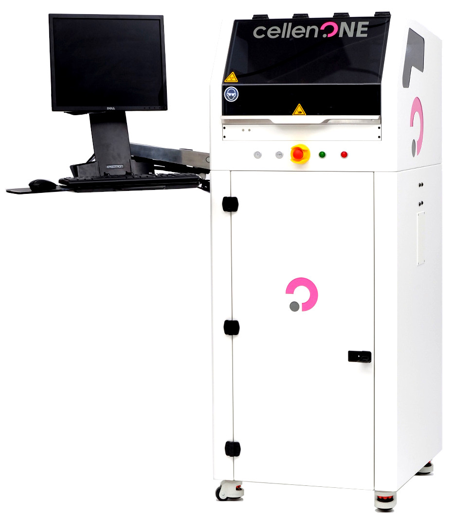
By addressing these bottlenecks — through improved microdissection tools, automation, and low-input sample prep protocols — spatial proteomics workflows are gradually becoming more robust and accessible. The cellenONE platform from Cellenion automates the entire single-cell isolation process, reducing the challenges of traditional methods and enabling greater efficiency without compromising accuracy.
Single-cell isolation
Integrating spatial proteomics with precise single-cell isolation enables researchers to connect molecular information with spatially defined cell populations, including rare or morphologically distinct cells. The cellenONE offers researchers the flexibility to isolate any number of cells per workflow, including single cells. Using image-based single-cell isolation combined with gentle acoustic aspirate/dispense technology, it ensures precise single-cell isolation, without contamination from neighboring cells, which is a crucial requirement when analyzing laser-microdissected or AI-identified targets in complex tissues. The system can be further customized based on cell size, shape, and fluorescence, enabling tailored isolation of specific phenotypes or tissue microenvironments.
Reduced reagent and sample use
The cellenONE system offers picoliter-range liquid dispensing, enabling streamlined single-cell proteomics workflows from a single platform. This is especially important when working with scarce clinical specimens or low-yield laser micro-dissections, as it reduces reagent waste and prevents the loss of irreplaceable samples. Built-in humidity controls and an oil overlay prevent evaporation and sample loss, while low input requirements enhance sensitivity and reliability from low-abundance samples.
Automation and reproducibility
The cellenONE proteomics workflows are fully automated, minimizing the need for manual handling and standardizing liquid dispensing and incubation steps to improve reproducibility. The system interfaces seamlessly with proteoCHIP consumable line, enabling complete sample preparation workflows to be completed in just three hours, compared to twelve hours using traditional methods. Additionally, samples can be transferred directly into the autosampler for seamless mass spectrometry analysis, streamlining the process from cell isolation to data acquisition.
Conclusion: Precision as the Foundation of Spatial Proteomics
Spatial proteomics is redefining our understanding of tissue heterogeneity, cellular microenvironments, and disease mechanisms by providing unprecedented spatial resolution in proteomic analysis. As workflows evolve, platforms like Cellenion’s cellenONE and proteoCHIP stand out, offering high-fidelity single-cell isolation, ultra-low reagent use, and seamless compatibility with downstream mass spectrometry. By addressing key challenges in sample preparation, Cellenion empowers researchers to unlock deeper insights with greater efficiency and precision, driving the future of spatial proteomics.
Related technologies: Cell sorting


