Discover


Webinar recording: Development And Optimization Of A 36-Color Panel For Deep Immunophenotyping
In this webinar, Cytek presented the development and optimization of a 36-color full spectrum flow cytometry panel for in-depth immunophenotyping of human T cells, with specific applications in adoptive cell therapy research.
June 19, 2026

Bioluminescence Distribution Of NPs In Rats’ Organs
Monitoring Nanoparticles distribution in rats' heart and liver with Alliance Chroma System
June 12, 2026


Quantitative Resolution Enhancement in In Vivo Two-Photon Intravital Imaging
Unlock high-resolution intravital imaging with advanced two-photon microscopy and computational enhancement. Visualize dynamic cellular processes in vivo with clarity, precision, and real-time insight.
May 18, 2026

Efficient DNA purification from cells: Automated workflows with sbeadex Lightning and CyBio FeliX
This application note presents a rapid, automated DNA purification workflow using the CyBio FeliX system with sbeadex Lightning technology, enabling high-quality results in just minutes while reducing hands-on time and increasing throughput.
May 6, 2026

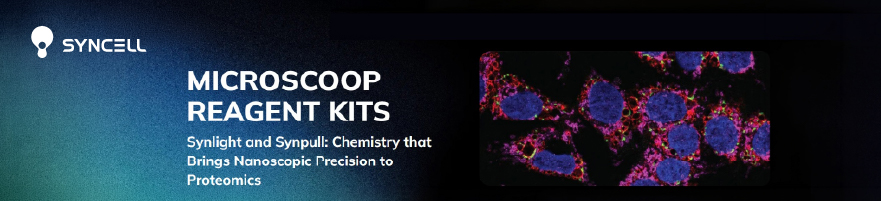
Introducing Synlight-Pure — Ultra-High Precision Photolabeling for Nanoscopic Proteomic Discovery
The webinar is demonstrating how microscopy-guided, patterned light activation enables protein purification directly from visually defined subcellular structures.
April 22, 2026
PHF19 drives the formation of PRC2 clusters to enhance motility in TNBC cells
This study investigates the subnuclear organization of the Polycomb repressive complex 2 (PRC2) in triple-negative breast cancer (TNBC) cells, combining in situ proteomics, high-resolution imaging, and functional genomics.
April 14, 2026
mRNA vaccine immunity is enhanced by hepatocyte detargeting and not dependent on dendritic cell expression
mRNA vaccine efficacy is enhanced by preventing expression in hepatocytes rather than promoting it in dendritic cells. The findings highlight tissue-specific targeting as a potential strategy for improving vaccines and cancer immunotherapies.
June 5, 2026
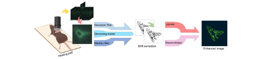
Combined Denoising and Resolution Enhancement Algorithms for Intravital Two-Photon Imaging
The study compared several image processing techniques, finding that the enhanced Super-Resolution Radial Fluctuations (eSRRF) combined with a trained denoising model significantly improved spatial resolution and mitochondrial structure visualization compared to other algorithms. Algorithms Evaluated: The study tested Gaussian and median filtering against a trained denoising model, followed by resolution enhancement using either eSRRF or […]
May 14, 2026
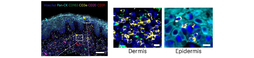
Cutaneous T cell lymphoma atlas reveals malignant TH2 cells supported by a B cell-rich tumor microen
The study provides a high-resolution atlas of cutaneous T cell lymphoma, identifying that malignant cells resemble TH2 cells. It highlights how a B cell-rich microenvironment supports tumor growth, offering new targets for therapy.
May 4, 2026

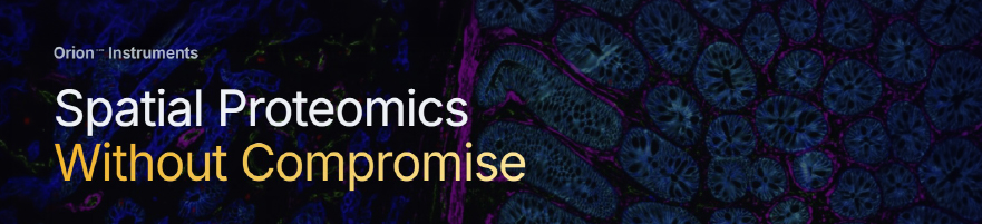
Introducing Orion HT
The New Standard for Spatial Biomarker Development in Translational and Clinical Research
April 20, 2026

Comprehensive analysis of B cell repopulation in ocrelizumab-treated patients with MS
Using the Curiox Pluto platform to automate immune profiling, ocrelizumab’s effects in MS reveal repopulation of transitional naïve B cells at 6 months and linked shifts in T/myeloid markers and plasma proteins.
April 7, 2026


A combination of systemic nanoparticles for caveolae-mediated gene delivery to the brain
This study shows that systemic mannitol induces caveolae in the brain endothelium. Mannitol-modified nanoparticles exploit this to cross the BBB, enhancing gene delivery.
May 20, 2026

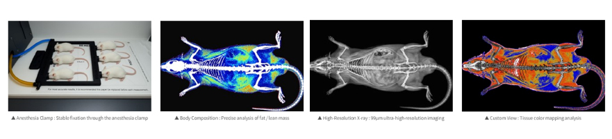
New product launch: InAlyzer2, Medikors
Next generation DXA Precision Analysis Technology
May 11, 2026
High-Throughput UVA-Based Thermal Shift Assays for Hydrophobic Proteins Using the qTOWER iris
This note demonstrates a UVA-based Thermal Shift Assay on the qTOWER iris, enabling sensitive detection of protein stability and expanding qPCR use to protein analysis.
April 23, 2026

Efficient DNA purification from cells: Automated workflows with sbeadex Lightning and CyBio FeliX
This application note presents a rapid, automated DNA purification workflow using the CyBio FeliX system with sbeadex Lightning technology, enabling high-quality results in just minutes while reducing hands-on time and increasing throughput.
April 16, 2026