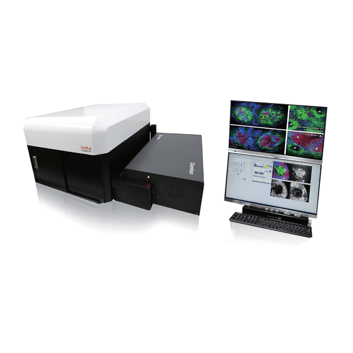


IntraVital Microscopy is a technique that enables you to directly observe the movement of live cells that make up living tissue in vivo.
Accelerate to discover
Back to filter
Related topics


Intravital imaging reveals the early astrocytic response to ischemic stroke
The use of IVIM-C intravital confocal microscopy provided a major advantage by enabling dynamic imaging of the same brain region in living animals over time. This allowed the researchers to capture the sequence of pathological events following ischemic stroke rather than relying only on endpoint histological analysis.
IVIM imaging revealed a rapid and transient increase in astrocytic H₂O₂ immediately after thrombosis, occurring within minutes and preceding the subsequent structural and molecular changes. The system also enabled longitudinal monitoring of vascular disruption, astrocyte responses and glial barrier formation, with imaging performed at the same location over several weeks.
These observations helped establish the temporal relationship between the early oxidative-stress response and later collagen deposition, glial barrier formation and neuronal loss. The study therefore demonstrates the value of IVIM technology for non-terminal, longitudinal visualization of disease progression and cellular dynamics in vivo, providing information that would be difficult to obtain from conventional fixed-tissue imaging alone.
Related technologies: IntraVital Microscopy