
Systems, which drive convergence of dynamic proteomics and single cell biology for the first time, are creating this deeper connection to accelerate curative medicines.
Accelerate to discover
Back to filter
Related topics
Perfusion: The Beating Heart of Single-Cell Biology and Cancer Immunotherapy
Single-cell biology has transformed our ability to uncover cellular diversity and dynamic behavior within complex tissues that are otherwise masked in population-level analyses. Yet, the precision of single-cell data critically depends on how cells are maintained and stimulated during experiments.
In static cultures, nutrient depletion and waste accumulation occur rapidly, altering cellular metabolism and gene expression. Perfusion – the continuous delivery and exchange of medium – has emerged as a key enabling technology for preserving cellular homeostasis and experimental reproducibility in single-cell systems. Perfusion overcomes these issues of static cultures by sustaining dynamic equilibrium, constantly delivering nutrients, removing waste, and maintaining optimal pH, oxygen, and osmotic balance.
Imagine you’re at a concert in a stadium. You’re in the front row in the pit, yet encapsulated in a crowd, trying to breathe, think, hungry and thirsty – but still trying to enjoy the music. Then someone pulls you out of the crowd, takes a picture and makes you give a review of the concert.
Now imagine instead you’re in a luxury box, with plenty of elbow room, being provided with refreshments and all the amenities – getting VIP service. Now, your interviewer does a Teams meeting with you, leaving your environment undisturbed, to obtain a more accurate review of the concert.
Good vs. Bad Perfusion
That, in the world of cell biology, is the difference perfusion can make – especially when you’re interrogating single, ex vivo or in vitro cells under the microscope. Perfusion maintains your subject in their best light.
The Science Behind the Flow
Cells are sensitive entities – even small shifts in pH, oxygen, glucose, or waste buildup can change their behavior. Just like humans don’t do well in a room full of carbon dioxide and dirt, cells don’t do well in stagnant media.
Encapsulation of single cells has come a long way in both therapy and research, but current methods still struggle to not only maintain cells viably for longer than a few hours (Heyman et al, 2021), and manifesting their full functional potential, but also to be able to assay these same cells without disrupting their equilibrium. For longer-term culture, even if a single cell is successfully captured in a droplet based on the Poisson distribution – or non-single-cell droplets are discarded – and after overcoming shear stress through a 30uM nozzle, the only current strategy to dilute waste and stress response proteins, and replenish nutrients, is to increase droplet size. However, this not only dilutes the concentration of secreted products but may also impair the optimal focal depth for the observer – and does this truly reverse the negative effects?
Perfusion systems keep the microenvironment replenished and stable. It ensures enhanced viability and supports proliferation, as well as a more physiologically relevant environment, reducing the need for manual intervention and improving the handling of sensitive cell types. And constant nutrient exchange results in happier, healthier, more predictable cells. More predictable cells lead to better biological data.
This is most important for functional screens where timing and kinetics matter — like identifying which cell secretes cytokines first or how long a killing event takes — by keeping the system in balance, you can get real-time data that actually reflects the biology, not just cells’ stress responses to a polluted, shrinking environment .
Perfusion also lets you integrate multiple assays, sequentially and/or concurrently, and allows for the rapid and precise application of reagents (such as drugs or signaling molecules), to individual cells in a dose-dependent and time-controlled manner. This is critical for studying dynamic cellular responses and pathway signaling with high precision.
Bringing Biology Closer to Physiology with Perfusion
Continuous media flow mimics in vivo conditions, enabling more accurate modeling of cell function, communication, and response within dynamic, multi-cellular systems.
Recreating the Tumor Microenvironment
Perfused microfluidic systems allow precise control over oxygen, nutrient, and cytokine gradients, closely mimicking in vivo conditions. This enables realistic modeling of tumor–immune interactions and infiltration dynamics (Zervantonakis et al., 2012; Businaro et al., 2013, Zenga et al., 2024)
Single-Cell Functional Profiling of Immune Responses
Using perfusion-based assays, researchers can monitor immune effector cell function — including CAR-T or NK cell cytotoxicity — in real time at single-cell resolution. These platforms reveal heterogeneity in killing efficiency and cytokine secretion that static cultures often miss (Varadarajan et al., 2012; Eyer et al., 2019, “Unravel the Complexity of Single Natural Killer Cell Cytotoxicity and ADCC Mediation”, “Phase I/II Study of Adaptive Manufactured Lentiviral Anti-CD20/Anti-CD19 Chimeric Antigen Receptor T Cells for Relapsed, Refractory Mantle Cell Lymphoma” ).
Dynamic Drug Screening and Resistance Modeling
Perfusion platforms simulate realistic pharmacokinetic exposure to checkpoint inhibitors, cytokines, or small molecules. This dynamic approach uncovers adaptive resistance mechanisms and helps identify optimal dosing strategies for immunotherapies (Dura et al., 2022).
Integrating Functional and Molecular Insights
By coupling perfusion assays with single-cell omics, researchers can link dynamic functional phenotypes – including long-term assays such as T-cell exhaustion or reactivation — to transcriptional programs within the same cells (Jang et al., 2021, Zenga et al., 2024). This integration accelerates biomarker discovery and rational design of next-generation immunotherapies.
High-Quality Functional Single-Cell Data Through Perfusion on the Beacon Optofluidic System
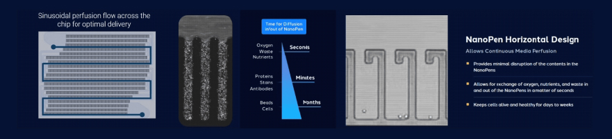
Here at Bruker Cellular Analysis, our Beacon Optofluidic system supports perfusion and, subsequently, exceptional functional single-cell data by using Opto-Electrical Positioning (OEP) to gently isolate cells in their own small NanoPen® chambers that are continuously perfused with fresh media at a rate of 1ml per day, facilitating long-term culture and real-time monitoring of individual cell behaviors and secreted molecules.
This light-driven platform allows for precise cell manipulation and real-time, functional, multi-parameter analysis of live cells, providing high-resolution, dynamic data across thousands of cells simultaneously for applications like antibody discovery, cell line development, and cell therapy research.
What’s even better: after their “stay” in the NanoPen chambers, cells can be recovered for further downstream work – apart from the effects of the passage of time, they are relatively unchanged from the moment they entered.
Final Thoughts
Perfusion represents more than a technical refinement – it is a fundamental enabler of reproducible, physiologically meaningful single-cell experimentation.
No one wants to make therapeutic decisions based on stressed-out cells acting uncharacteristically. By maintaining homeostatic conditions and enabling controlled perturbation, perfusion ensures that the data we extract from single cells reflect genuine biology rather than artifacts of culture. As single-cell systems continue to mature, the principles of perfusion will remain central to achieving quantitative, dynamic, and integrative insights into cellular behavior.
Related technologies: Functional biology