Discover


Bioluminescence Distribution Of NPs In Rats’ Organs
Monitoring Nanoparticles distribution in rats' heart and liver with Alliance Chroma System
June 12, 2026

mRNA vaccine immunity is enhanced by hepatocyte detargeting and not dependent on dendritic cell expression
mRNA vaccine efficacy is enhanced by preventing expression in hepatocytes rather than promoting it in dendritic cells. The findings highlight tissue-specific targeting as a potential strategy for improving vaccines and cancer immunotherapies.
June 5, 2026

Efficient DNA purification from cells: Automated workflows with sbeadex Lightning and CyBio FeliX
This application note presents a rapid, automated DNA purification workflow using the CyBio FeliX system with sbeadex Lightning technology, enabling high-quality results in just minutes while reducing hands-on time and increasing throughput.
April 16, 2026


Accelerating Antibody Discovery with the WOLF G2 Cell Sorter
Site-directed antibodies targeting driver mutations of the KRAS protein
March 16, 2026


Introducing IVM-FS: Real-Time Cellular Insightsinto Rats and Other Medium-Sized Animals
Rats are increasingly valued for intravital imaging due to their larger body size and organ systems, which enable more precise surgical preparation, stable window implantation, and repeated long-term imaging - advantages over mice.
March 3, 2026

Harnessing the WOLF Cell Sorter for Cutting‑Edge Senescence Research
Senescent Endothelial Cells in Cerebral Microcirculation Are Key Drivers of Age-Related Blood–Brain Barrier Disruption, Microvascular Rarefaction, and Neurovascular Coupling Impairment in Mice
February 18, 2026

Efficient DNA purification from cells: Automated workflows with sbeadex Lightning and CyBio FeliX
This application note presents a rapid, automated DNA purification workflow using the CyBio FeliX system with sbeadex Lightning technology, enabling high-quality results in just minutes while reducing hands-on time and increasing throughput.
May 6, 2026

PHF19 drives the formation of PRC2 clusters to enhance motility in TNBC cells
This study investigates the subnuclear organization of the Polycomb repressive complex 2 (PRC2) in triple-negative breast cancer (TNBC) cells, combining in situ proteomics, high-resolution imaging, and functional genomics.
April 14, 2026


Whole genome analysis and in vivo safety assessment of probiotic candidate Lactobacillus acidophilus
Probiotics, particularly strains of lactic acid bacteria (LAB), are recognized for their beneficial effects on gut health. Lactobacillus acidophilus L177, a promising probiotic strain isolated from canine feces, was evaluated for safety and probiotic.
March 10, 2026
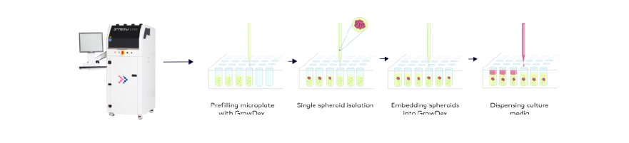
Combining GrowDex and spheroONE for automated and highly reproducible 3D cell culture workflows
This application note demonstrates the compatibility of spheroONE and GrowDex to automate 3D cell culture workflows. spheroONE is a nanoliter dispenser enabling sorting and isolation of spheroids and organoids.
February 25, 2026

Use of Vizgen’s MERSCOPE Platform to Uncover New Insights about Aging
Microglia activation orchestrates CXCL10-mediated CD8+ T cell recruitment to promote aging-related white matter degeneration - peer-reviewed paper outlines aging-related changes in white matter by single-cell and spatial transcriptomics
February 11, 2026

High-Throughput UVA-Based Thermal Shift Assays for Hydrophobic Proteins Using the qTOWER iris
This note demonstrates a UVA-based Thermal Shift Assay on the qTOWER iris, enabling sensitive detection of protein stability and expanding qPCR use to protein analysis.
April 23, 2026

Standardized Thawing and Sample Preparation of ARCTis Cryopreserved Human Tumor Cells
The standardized and automatic handling of cryopreserved biological samples plays an increasingly important role in today’s in vitro testing. This is due to the trend to develop more complex disease models to increase the data quality of studies.
March 30, 2026


Preclinical Evaluation of 223RaCL2 and Immune Checkpoint Inhibitors in Prostate Cancer Bone Mets
This study investigates the therapeutic and immunological effects of combining Radium-223 dichloride (Xofigo) with immune checkpoint inhibitors (ICIs) in a clinically relevant, immunocompetent murine model of prostate cancer bone metastasis.
March 9, 2026

Comparing Microscoop with Proximity Labeling Techniques APEX2 and BioID
Discover how Microscoop, APEX2, and BioID offer unique advantages for mapping protein-protein interactions—whether in fixed tissue or live cells. Learn which method fits your proteomics research best!
February 23, 2026