Products
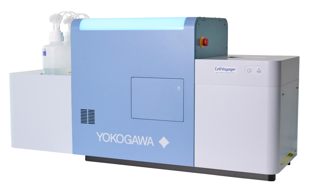
CQ3000 – Yokogawa
High-definition 3D microscopic imaging of live cell cultures
High-Content Analysis System, featuring advanced assays in environments closer to the human body such as organoids—which replicate certain structures and functions of human organs— or organ-on-a-chip models that mimic physiological functions of organs on microfluidic chips.
In addition, multicolor imaging using multiple fluorescent dyes allows complex assays like morphological profiling of cellular components and organelles through techniques such as Cell Painting.
Key Features:
High Resolution
・High-NA water-immersion objectives provide bright, deep imaging
・Equipped with up to 2 high–quantum-efficiency sCMOS cameras
・Uniformizer enables even illumination across the entire field of view
High Throughput
・Proprietary confocal method enables rapid acquisition of 3D images
・Second camera allows simultaneous dual-wavelength imaging
・Optional ultra-high-speed mode up to 100 fps is ideal for fast applications such as cardiomyocyte beating and calcium signaling
Live-Cell Imaging
・Industry-leading incubator performance ensures stable long-term live-cell imaging
・Supports incubation for up to 7 days
・Confocal scanner unit CSU enables imaging with low photobleaching and low phototoxicity
Automation
・Automatically scans the entire sample and captures images that meet predefined conditions
・Supports external system integration through an API
・Outputs data in widely compatible formats such as OME-TIFF and OMERO
More info at: CellVoyager High-Content Analysis System CQ3000 | Yokogawa Europe
Documentation