Discover


A combination of systemic nanoparticles for caveolae-mediated gene delivery to the brain
This study shows that systemic mannitol induces caveolae in the brain endothelium. Mannitol-modified nanoparticles exploit this to cross the BBB, enhancing gene delivery.
May 20, 2026


Quantitative Resolution Enhancement in In Vivo Two-Photon Intravital Imaging
Unlock high-resolution intravital imaging with advanced two-photon microscopy and computational enhancement. Visualize dynamic cellular processes in vivo with clarity, precision, and real-time insight.
May 18, 2026

CXCL12+ fibroblastic reticular cells in lymph nodes facilitate immune tolerance
The popliteal LNs were exposed surgically and confocal intravital imaging was performed (IVM-CMS3). Twenty-five Z-stack images were obtained with a 3 μm axial interval. Time-lapse images were obtained at a 1-minute time interval for 20 minutes.
January 26, 2026

Reanalyze your in-vivo optical images with Aura Software
Aura is a free license software application produced by Spectral Instruments Imaging, a Bruker Company.
October 23, 2025

Optimizing Substrate Dosing for ReliableBioluminescence Imaging (BLI)
Bioluminescence imaging relies on precise D-luciferin administration, as dosing variability impacts signal intensity and data consistency. Standardizing substrate delivery is essential for reproducible and high-quality in vivo imaging results.
September 18, 2025


Curious how PET imaging and reporter gene tracking are transforming CAR-T cell therapy in oncology?
Recorded webinar is recommended if you are working or you are interested by translational studies in oncology, immune therapy, and biomedical imaging.
June 2, 2025
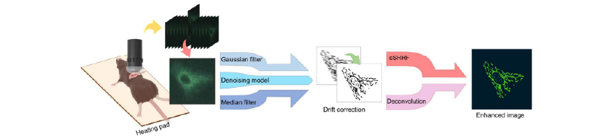
Combined Denoising and Resolution Enhancement Algorithms for Intravital Two-Photon Imaging
The study compared several image processing techniques, finding that the enhanced Super-Resolution Radial Fluctuations (eSRRF) combined with a trained denoising model significantly improved spatial resolution and mitochondrial structure visualization compared to other algorithms. Algorithms Evaluated: The study tested Gaussian and median filtering against a trained denoising model, followed by resolution enhancement using either eSRRF or […]
May 14, 2026

Combination of low-energy X-ray & citric acid to inactivate E.coli, S.Typhimurium, L.Monocitogenes
Combined treatment with 0.5 kGy X-ray and 0.1 % CA significantly decreased biofilm cell counts by 5.10, 4.31, and 3.96 log CFU/coupon for E. coli O157:H7, S. Typhimurium, and L. Monocytogenes, respectively.
December 3, 2025

Can you solve mouse CT imaging with 50um spatial resolution ?
MOLECUBES, a Bruker Company offers small footprint microCT for mice and rats, which meets most of your needs for in-vivo analysis and doesn't sacrifice the budget.
October 22, 2025

Subcutaneous delivery of MSC induces immunoregulatory effects in the lymph node
Mice were imaged under anaesthesia (2.5% isoflurane in oxygen), daily until the bioluminescence signal disappeared. Imaging was performed in an Ami-HTX Small Animal Imager and analysed with Aura Imaging Software, from Spectral Instruments Imaging.
September 12, 2025
High-Resolution Intravital In Vivo Imaging with AI-Powered Image Denoising
In this webinar, experts discuss cutting-edge advancements in microscopy imaging and denoising technologies.
April 14, 2025

New product launch: InAlyzer2, Medikors
Next generation DXA Precision Analysis Technology
May 11, 2026

Development of new albumin‑binding radiotracers for PET imaging of CSF
In this study by Peltoniemi et al., PET/CT imaging using the X-CUBE (CT) and ß-CUBE (PET) was used to non-invasively visualize cerebrospinal fluid (CSF) flow and glymphatic system dynamics in rats.
November 28, 2025

Elevating Intravital Imaging with AI: Introducing the AI-Image Denoiser
AI-Image Denoiser, our best-in-class, AI-powered image enhancement software designed specifically for high-speed intravital microscopy.
October 21, 2025

Pericytes promote metastasis by regulating tumor local vascular tone and hemodynamics
The anesthetized mouse was placed on a customized stage for tumor immobilization and colon tumor was exposed to intravital microscopy (IVM-MS2, IVIM Technology, Korea) equipped with 25× water immersion objective.
September 10, 2025