Discover


A combination of systemic nanoparticles for caveolae-mediated gene delivery to the brain
This study shows that systemic mannitol induces caveolae in the brain endothelium. Mannitol-modified nanoparticles exploit this to cross the BBB, enhancing gene delivery.
May 20, 2026

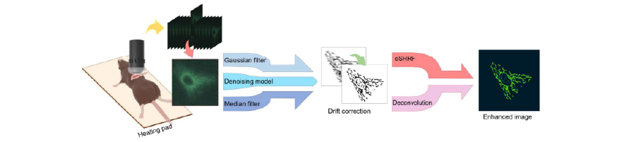
Combined Denoising and Resolution Enhancement Algorithms for Intravital Two-Photon Imaging
The study compared several image processing techniques, finding that the enhanced Super-Resolution Radial Fluctuations (eSRRF) combined with a trained denoising model significantly improved spatial resolution and mitochondrial structure visualization compared to other algorithms. Algorithms Evaluated: The study tested Gaussian and median filtering against a trained denoising model, followed by resolution enhancement using either eSRRF or […]
May 14, 2026

High-Throughput UVA-Based Thermal Shift Assays for Hydrophobic Proteins Using the qTOWER iris
This note demonstrates a UVA-based Thermal Shift Assay on the qTOWER iris, enabling sensitive detection of protein stability and expanding qPCR use to protein analysis.
April 23, 2026

Comprehensive analysis of B cell repopulation in ocrelizumab-treated patients with MS
Using the Curiox Pluto platform to automate immune profiling, ocrelizumab’s effects in MS reveal repopulation of transitional naïve B cells at 6 months and linked shifts in T/myeloid markers and plasma proteins.
April 7, 2026


Accelerating Antibody Discovery with the WOLF G2 Cell Sorter
Site-directed antibodies targeting driver mutations of the KRAS protein
March 16, 2026

Harnessing the WOLF Cell Sorter for Cutting‑Edge Senescence Research
Senescent Endothelial Cells in Cerebral Microcirculation Are Key Drivers of Age-Related Blood–Brain Barrier Disruption, Microvascular Rarefaction, and Neurovascular Coupling Impairment in Mice
February 18, 2026

Efficient DNA purification from cells: Automated workflows with sbeadex Lightning and CyBio FeliX
This application note presents a rapid, automated DNA purification workflow using the CyBio FeliX system with sbeadex Lightning technology, enabling high-quality results in just minutes while reducing hands-on time and increasing throughput.
May 6, 2026

Efficient DNA purification from cells: Automated workflows with sbeadex Lightning and CyBio FeliX
This application note presents a rapid, automated DNA purification workflow using the CyBio FeliX system with sbeadex Lightning technology, enabling high-quality results in just minutes while reducing hands-on time and increasing throughput.
April 16, 2026
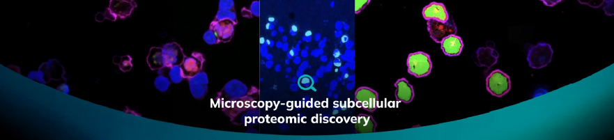
Microscopy-guided subcellular proteomic discovery by high-speed ultra-content photo-biotinylation
Syncell developed a method combining microscopy, photo-biotinylation, and mass spectrometry, enabling high-resolution mapping of proteins in subcellular regions, accelerating discoveries in spatial proteomics research.
March 23, 2026

NEW: TIDeX – Total Ionizing Dose Instrument
The all-in-one X-ray platform designed to streamline Total Ionizing Dose (TID) effect testing with accuracy, control, and repeatability
March 12, 2026

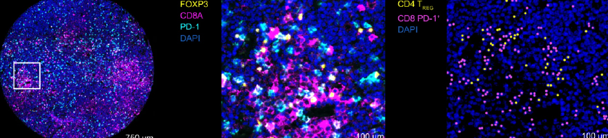
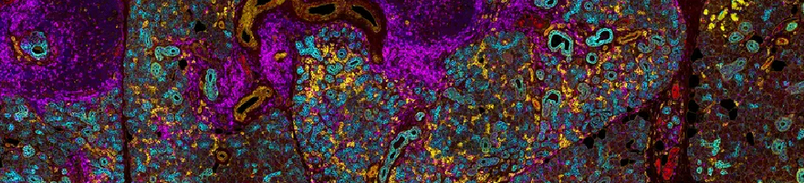
Integrative multi-omics reveals a regulatory and exhausted T-cell landscape in CLL
T-cell exhaustion contributes to immunotherapy failure in chronic lymphocytic leukemia (CLL). T cells from CLL patients' blood, bone marrow, and lymph nodes were analyzed using single-cell RNA sequencing, mass cytometry, and tissue imaging.
February 12, 2026
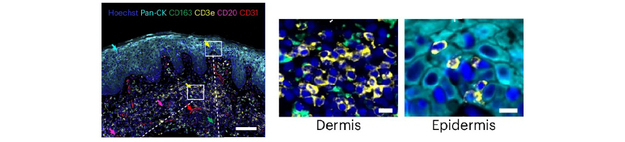
Cutaneous T cell lymphoma atlas reveals malignant TH2 cells supported by a B cell-rich tumor microen
The study provides a high-resolution atlas of cutaneous T cell lymphoma, identifying that malignant cells resemble TH2 cells. It highlights how a B cell-rich microenvironment supports tumor growth, offering new targets for therapy.
May 4, 2026

PHF19 drives the formation of PRC2 clusters to enhance motility in TNBC cells
This study investigates the subnuclear organization of the Polycomb repressive complex 2 (PRC2) in triple-negative breast cancer (TNBC) cells, combining in situ proteomics, high-resolution imaging, and functional genomics.
April 14, 2026
Molecular and Spatial Drivers of Immunotherapy Response in Mucosal Melanoma
Findings suggest that distinct genomic features, tumor states, and microenvironments differentiate ICB response and progression in MM, highlighting key tumor-intrinsic and microenvironmental factors associated with prognosis.
March 18, 2026

Preclinical Evaluation of 223RaCL2 and Immune Checkpoint Inhibitors in Prostate Cancer Bone Mets
This study investigates the therapeutic and immunological effects of combining Radium-223 dichloride (Xofigo) with immune checkpoint inhibitors (ICIs) in a clinically relevant, immunocompetent murine model of prostate cancer bone metastasis.
March 9, 2026